MENU

Optomap Retinal Imaging

Retinal imaging aids in the early detection of disease, which is essential to reduce the risk to your sight and health

Retinal imaging aids in the early detection of disease, which is essential to reduce the risk to your sight and healthLocated in the back of your eye, your retina is the only place in your body where blood vessels can be seen directly. These blood vessels can provide insight on diseases and conditions the body may be experiencing. By looking into the back of your eye, Dr. Miller can find early signs of conditions such as stroke, heart disease, hypertension, and diabetes.

Eye doctors have been examining the retinas for years to aid in the early diagnosis of these conditions, but old technology only allowed them to view a small window of the back of the eye.

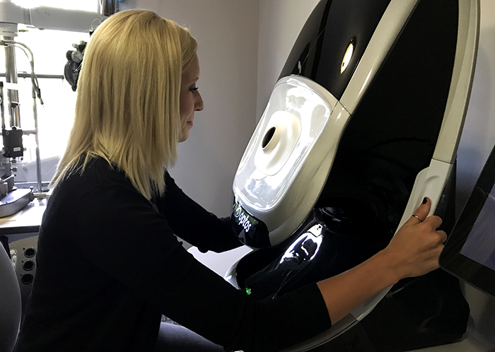
At Visual Eyes, we’ve invested in the latest technology that gives us an ultra wide angle view of the retina. This allows us to catch conditions when they begin to appear on your retina, long before they cause changes to vision or pain.

By utilizing Optomap retinal Imaging, we able to quickly take a picture of your retina and discuss any findings with you in just a few minutes.

An Optomap image is taken quickly and painlessly

When you come in for your annual eye exam, our team will take an Optomap image of your retina. The process is fast, painless, and completely comfortable. Nothing touches your eye at any time and Optomap is suitable for the whole family. Simply look into the device one eye at a time, and wait for the flash of light, which lets you know the image has been taken. It’s similar to looking through a keyhole. For most patients, this process can be done without dilation drops. Dr. Miller will decide if your pupils will need to be dilated depending on your eyes. In all, the image capture takes less than a second and images are available immediately for review. You will be able to see your own retina!

Traditional methods of retina imaging left the peripheral vision out

The ultra-widefield retinal image the Optomap takes captures more than 80 percent of the retina in one panoramic image. Traditional imaging typically only shows 15 percent at a time. This used to mean that early signs of disease that were present in the periphery of you retina could be missed and remain undetected. But, with the Optomap, Dr. Miller can see 200º of the retina, instead of only 45º.

The Benefits of an Optomap:

  • By mapping your eyes we can facilitate early protection from vision impairment or blindness
  • Check for early detection of life-threatening diseases like cancer, stroke, and cardiovascular disease
To learn more about what we check for, visit our comprehensive eye exam page.